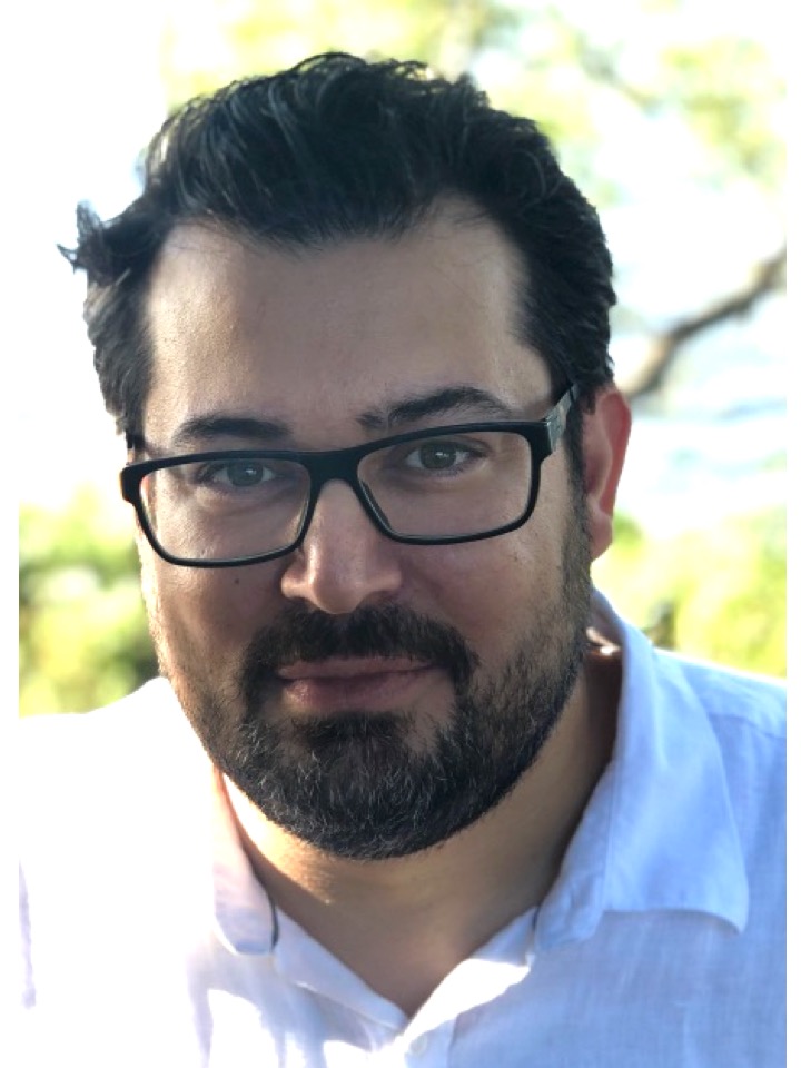
Alexander Marneros, M.D., Ph.D.
|
Clinicn Investigator, Assc Prf Dermatology Research MGH, Mass General Research Institute |
|
Associate Professor of Dermatology Harvard Medical School |
|
Associate Dermatologist Dermatology, Massachusetts General Hospital |
| PhD Harvard Medical School 2003 |
| MD University of Cologne School of Medicine 2003 |
Research Interests
Research Narrative
Our laboratory is interested in mechanisms that affect wound healing, inflammation, angiogenesis, epithelial differentiation and fibrosis.
Project 1: Abnormal Angiogenesis During Aging: Age-Related Macular Degeneration
Abnormal blood vessel growth is an important component in the pathogenesis of various common diseases. Pathological angiogenesis is stimulated by an inflammatory response that involves many different cell types, including proangiogenic macrophages. Our laboratory focuses on the factors and cell types that promote pathologic angiogenesis in development, in the adult and during aging.
Age-related macular degeneration (AMD) is the most common cause of irreversible blindness in the elderly. Non-exudative ("dry") AMD manifests with degeneration of the retinal pigment epithelium (RPE) and sub-RPE deposit formation with progressive age. Neovascular ("wet") AMD manifests with pathologic neovascularization from the choroid into the retina. Often both forms co-occur, suggesting a common pathomechanism.
We have recently shown in a new genetic mouse model of AMD that increased VEGF-A is sufficient to cause both forms of AMD, providing evidence for a unifying pathomechanism in advanced AMD. In this model, proangiogenic macrophages stimulate retinal glia cells to promote choroidal neovascularization (Cell Reports, 2013; FASEB J , 2014; FASEB J, 2018).
Furthermore, we could show that the NLRP3 inflammasome promotes VEGF-A-induced AMD pathologies, and targeting inflammasome components inhibits these pathologies (EMBO Mol Med, 2016). Our work focuses on elucidating the mechanisms that regulate molecular pathways and cellular interactions during AMD pathogenesis, with the aim of identifying novel therapeutic approaches for patients with AMD.
Project 2: Role of macrophages in angiogenesis and the wound healing response
Multiple cell types interact in a highly coordinated fashion in response to injury to promote wound healing. Abnormalities in this complex process can result in exuberant wound angiogenesis or scarring.
We use in vivo models of wound healing to determine the spatiotemporal contributions of various cell types in this process. In a laser-injury model of choroidal neovascularization and in skin wound healing models, we could show that macrophages become alternatively activated (M2-type) and promote wound angiogenesis, in part by stimulating retinal glia cells to express proangiogenic growth factors. Specific ablation of macrophages inhibits the early wound healing response to injury and blocks wound angiogenesis (Am J Pathol, 2013, 2016; Cell Reports, 2013).
Our current research investigates which signaling pathways drive polarization of macrophages to the proangiogenic M2-type in vivo and how this polarization can be inhibited (JBC, 2014). Overall, we aim to define the contributions of individual inflammatory cell populations to wound healing and neovascularization. Furthermore, we aim to develop targeted therapies that specifically block M2-type macrophages.
Project 3: Regulaton of skin formation during development: causes of congenital wounds
Wound healing and wound angiogenesis occur in response to injury. However, congenital wounds are observed in various genetic syndromes as a manifestation of a skin morphogenesis defect during embryonic development. Identifying the genetic basis for these conditions and syndromes promises to identify novel genes and pathways that are critical for proper skin formation and whose impairment results in wounds.
A prototypical congenital skin disease that manifests with a wound present at birth is aplasia cutis congenita (ACC). Using a combination of genome-wide linkage analysis and exome sequencing we recently identified the causative mutation for ACC (PLOS Genetics, 2013; Am J Hum Genet., 2013; JID, 2015). We found that the ribosomal GTPase BMS1 is mutated in ACC, which results in a maturation defect of the small ribosomal subunit and leads to a nucleolar stress response with a p21-mediated G1/S phase transition delay and a reduced cell proliferation rate. Global proteomic and transcriptional analyses revealed a central role of p21 activation for the ACC phenotype. Our laboratory investigates how a mutation in a ubiquitous ribosomal protein leads to localized skin defects without affecting other organ systems. We seek to obtain a comprehensive understanding of the disease mechanisms of congenital wound healing disorders.
Project 4: Epithelial differentiation mechanisms in the kidney: role for chronic kidney disease, renal fibrosis and cyst formation
Our studies on epidermal morphogenesis in the skin have led us to assess molecular mechanisms that control epithelial differentiation in other epithelial tissues, particularly the kidney. We identified novel regulators of the epithelial differentiation process of the nephron, which have previously not been considered to affect kidney function. Using mouse genetics approaches we are aiming to define the molecular mechanisms that control epithelial differentiation in the kidney during development and in the adult. We also aim to determine how epithelial defects in the kidney may contribute to renal fibrosis and cyst formation.
| amarneros@mgh.harvard.edu |
| 6176437170 |
|
Cutaneous Biology Research Center CNY-Building #149 149 13th Street Charlestown, MA 02129-2000 |
