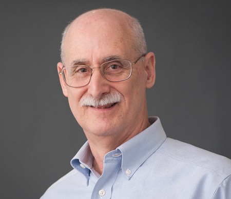
Jerome Ackerman, Ph.D.
|
Investigator, Assoc Prof (M) Rad Rsch MART 1 MO NE, Mass General Research Institute |
|
Associate Professor of Radiology Harvard Medical School |
| B.S., Chemistry Stony Brook University 1971 |
| Ph.D., Physical Chemistry Massachusetts Institute of Technology 1976 |
| Postdoctoral University of California, Berkeley 1977 |
Research Interests
Research Narrative
In the Biomaterials and Extremity Scanner Laboratory we develop magnetic resonance instrumentation and methods for interventional radiology; bone and synthetic biomaterials; and other novel applications.
MRI Therapy:
Our MR Radiofrequency Ablation technology permits the use of the MRI scanner as a therapeutic tool by enabling tumor ablations to be performed with RF heating using the scanner as the energy source. By extending this technology in a novel method called MR Coagulation, we coagulate intravascularly delivered biomaterials using scanner-derived RF energy to repair vascular defects. In both cases, the MRI scanner serves a dual ("theranostic") purpose as 1) a diagnostic tool to guide the intervention and monitor its progress and outcome, and 2) a therapeutic tool that delivers the energy to accomplish the therapy.
Solid State MRI and MRS of Bone, Arterial Calcification, and Biomaterials:
We are developing a compact, low cost MRI scanner for extremities (arms and legs) that can be sited anywhere. Among our research interesst are the compositional characterization of bone during growth and maturation, and quantitative solid state MR imaging of bone mineral and matrix to characterize metabolic bone disease. We use high field solid state cross polarization/magic angle spinning NMR spectroscopy to study vascular calcification in atherosclerotic plaque and heart valves. Synthetic biomaterials of interest include polymers, ceramics and composites.
Moving MRI:
We are developing an MRI scanner that operates while the entire magnet and the subjectt are in motion during scanning. This has applications to the study of the vestibular system (perception of bodily orientation and motion) and traumatic brain injury (displacement of brain tissue and fluids during acceleration).
| jlackerman@mgh.harvard.edu |
| 6177263083 |
|
Athinoula A. Martinos Center for Biomedical Imaging CNY-Building #149 149 13th Street 2-2.320 Charlestown, MA 02129 |
