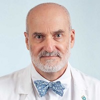
Matthew Frosch, M.D., Ph.D.
|
Clinicn Investigator, Full Prf PATHM CP FAC, Mass General Research Institute |
|
Admissions Chair, HST Harvard Medical School |
|
Lawrence J. Henderson Professor of Pathology and Health Sciences and Technology Harvard Medical School |
|
Pathologist Pathology, Massachusetts General Hospital |
| MD Harvard Medical School 1987 |
| PhD Harvard Medical School 1987 |
Research Interests
Research Narrative
My lab aims to understand cerebral amyloid angiopathy (CAA), using mouse models and human tissue. In this disease, the peptide Aβ deposits in the walls of blood vessels and is associated with risk of hemorrhage (‘lobar hemorrhages’). This peptide is the same material that forms the plaques of Alzheimer disease, and nearly all patients with Alzheimer disease have pathologic evidence of CAA as well. CAA also occurs in the absence of histologic evidence of Alzheimer disease, and can present with hemorrhages or with cognitive changes. In clinicopathologic studies, we have found that this latter presentation is associated with the presence of an inflammatory response, often containing giant cells. This subset of patients can have dramatic recoveries of cognitive function after immunosuppressive therapy.
We are interested in the sequence of events by which Aβ is deposited in blood vessels, what factors determine the distribution of involvement, what the consequences are for the cells of the vessel and how this material can respond to therapeutic interventions that have been shown to alter Aβ deposits in the brain (immunotherapy, gamma-secretase inhibitors). Current topics of particular interest involve the timing of oxidative stress induction by CAA, expression of matrix degrading enzymes such as MMP-9 and induction of apoptosis in vascular smooth muscle cells. We use serial in vivo multiphoton imaging with specific probes for these various processes and link the spatial and temporal distribution of the pathologic changes with the development of CAA. We complement these studies with immunohistochemistry, image reconstruction and laser capture microdissection to define alterations in gene expression that occur in smooth muscle cells in the proximity of amyloid deposits of CAA. Finally, our observations in mouse models of CAA can be validated through the use of human autopsy tissue, collected through the Massachusetts Alzheimer Disease Research Center Neuropathology Core.
I also work with a range of collaborators to understand the relationship between neuropathologic findings in the setting of disease – including Alzheimer disease, Parkinson disease, Amyotrophic Lateral Sclerosis and others – and other biochemical or functional markers of disease. These studies include advancing imaging methods (DTI, OCT and others) as well as various genetic studies (deep sequencing as well as GWAS), cell biology and structural biology.
| mfrosch@mgh.harvard.edu |
| 6177265156 |
|
Warren 275 Charles Street 334 Boston, MA 02114 |
